In eukaryotic transcriptomics, exons are traditionally conceptualized as stable coding blocks averaging 150 base pairs (bp) in humans. However, the discovery of micro-exons and their ultimate extreme - nano-exons - has challenged this definition. Nano-exons are exceptionally short coding sequences, often ranging from just 1 to 15 nucleotides.
Unlike standard exons, which provide large structural domains to a protein, nano-exons function as precision biochemical switches. Their alternative inclusion or exclusion inserts or deletes merely one to five amino acids within highly flexible loop regions. This subtle alteration selectively tunes protein-protein interactions, phosphorylation kinetics, or binding affinities without disrupting the global fold. Evolutionary analysis reveals that nano-exons are intensely conserved, exhibiting massive enrichment in the vertebrate nervous system, where they act as critical regulators of neurogenesis and synaptic plasticity.
Historically, nano-exons were virtually invisible. Standard short-read RNA-Seq detection relies on split-read alignment tools (e.g., STAR, HISAT2) to identify exon-exon junctions. When a read spans a nano-exon, the "anchor" sequence matching the tiny exon is too short (frequently < 5 bp), causing standard seed-and-extend heuristics to discard the alignment as random noise or a multi-mapping artifact.
Modern detection relies on specialized pipelines or alternative sequencing modalities:
- Hybrid Workflows (MicroExonator, OLego): These tools scan short-read data for identically soft-clipped or unmapped sequence overhangs at canonical splice boundaries to infer the presence of unannotated micro-exons.
- Long-Read Sequencing (PacBio, Oxford Nanopore): By capturing full-length continuous transcript isoforms, long reads natively preserve the full exon connectivity chain.
- Post-Processing Re-aligners (MisER): These algorithms scan long-read BAM files for localized clusters of insertion/mismatch errors near known splice sites and apply a specialized local alignment matrix with lowered gap-open penalties to resolve missing micro-exons.
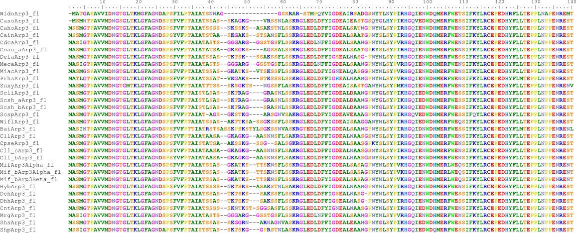
However, there are still cases where these methods fail. Consider a genomic architecture of a 4-bp nano-exon. See the figure for the example of the Arp3 gene of the yeast Babjeviella inositovora. The two competing processing paths yield two entirely distinct mature mRNAs:
- Case 1 (nano-exon skipped): The downstream GT bp is considered the donor site, treating the 4-bp as part of an extended exon. The mature mRNA reads: [Exon 1] + G + T + A + C + [Exon 2].
- Case 2 (nano-exon included): The upstream GT, the correct donor site, is used and the 4-bp nano-exon sequence added. The mature mRNA reads: [Exon 1] + A + C + A + A + [Exon 2].
Why is Case 2 correct? The amino acids around the and including the nano-exon are 100% conserved in all yeasts, and other yeasts have either (while not both) of the splice sites.
This precise sequence topology represents a catastrophic blind spot for standard bioinformatics. Because the sequences of the two potential transcripts are completely different but identical in length, typical length-profiling metrics fail. More critically, alignment algorithms are mathematically hardcoded to favor Case 1 over Case 2. To map a Case 2 long or short read correctly, an aligner must open two distinct introns. In any dynamic programming matrix (such as Smith-Waterman), opening an intron incurs a severe gap-open penalty (-G). The score reward for matching a mere 4 base pairs (max +4) is microscopically small. Consequently, the math always dictates that the aligner should open a single continuous intron and absorb the 4-bp difference as a cluster of 1 to 4 sequencing mismatches at the end of Exon 1.
Because the read bridges the entire gap, no soft-clipping occurs, completely blinding tools like MicroExonator. Concurrently, error-prone long reads mask these true junction mismatches within the background sequencing noise, causing tools like MisER to filter them out as random artifacts.
Can deep learning resolve these structural dead zones? In the human genome, the answer is increasingly yes. Context-aware AI models like SpliceAI leverage deep residual convolutional networks to scan up to 10,000 bp of flanking genomic sequence. Rather than scoring local motifs in isolation, these networks evaluate the macro-sequence topology: chromatin signatures, competitive splice-site kinetics, and hidden downstream/upstream Exonic or Intronic Splicing Enhancers (ESEs/ISEs). If a human 4-bp sequence possesses the explicit flanking binding code of specialized micro-exon assembly factors (such as Srrm4), SpliceAI will accurately predict the dual-splicing event, ignoring the raw alignment penalty limitations.
However, this AI capability dissolves when applied to far less studied species or species with compact genomes such as fungi. Vanilla SpliceAI is heavily bottlenecked by "human bias" - its 10kb context window assumes sprawling mammalian gene architecture. In a dense fungal genome, where introns are frequently <200 bp and intergenic spaces are minuscule, a 10kb window spans multiple neighboring genes, promoters, and termination signals. This dense cluster of unrelated biological signals completely disorients the network's competitive suppression layers, leading to massive oversuppression or an explosion of false positives.
Furthermore, fungal splicing relies on different regulatory codes. Without extensive, species-specific retraining data, which less studied organisms lack, deep learning models remain just as blind to these tiny sequence switches as the traditional heuristics that preceded them.